Over 13.9 million adults are affected by branch retinal vein occlusion (BRVO) globally.
Blood is carried throughout your body, including your eyes, through arteries and veins. One main artery and one main vein provide all the oxygen and nutrients for the retina of the eye.
Branch retinal vein occlusion (BRVO)
BRVO is a retinal disease which occurs when the branches of the retinal vein become blocked.
Blood and fluid leak out into the retina when a vein is blocked. This fluid might cause the macula to expand, compromising your central vision.
Without venous blood circulation from the retina, retinal disease will occur as the nerve cells die and eyesight deteriorates.
What causes BRVO
The majority of blockages resulting from BRVOs occur at an arteriovenous crossing, the place on the retina where a retinal artery and vein cross over each other.
At this cross-over point both vessels share a common sheath (connective tissue), and the vein becomes constricted when the artery loses flexibility due to atherosclerosis or hardening of the arteries.
The turbulent blood flow in the restricted vein encourages clotting, resulting in a blockage or occlusion. This obstruction prevents blood flow, resulting in fluid leaks in the macula (macular edema) and ischemia (low blood flow) in the blood vessels supplying the macula.
The common risk factors for BRVO are:
- Cardiovascular (heart) disease
- Glaucoma
- Being overweight or obese
- Uncontrolled high blood pressure
Symptoms of BRVO
BRVO produces a painless, rapid loss of vision. BRVO can go unnoticed, with no symptoms if the affected area is not in the center of the eye.
Visual floaters from a vitreous hemorrhage, blood vessels bleeding into the vitreous gel of the eye, might be the major symptom in rare situations of an undetected vein blockage; this is caused by the growth of abnormal new blood vessels in the retina.
SEE RELATED: Guide to Retinal Diseases
If you’ve experienced any vision loss contact an eye doctor near you.
How is BRVO diagnosed?
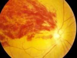
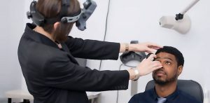
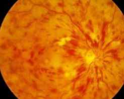
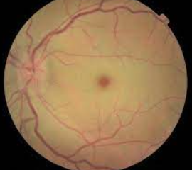
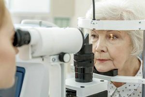
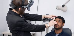
BRVO is most often diagnosed during an retinal eye exam with digital retinal imaging that reveals a retinal hemorrhage (blood vessels leaking into the retina), twisted and thickened blood vessels, and retinal edema (swelling with fluid).
Two types of in-depth retinal imaging tests aid the diagnosis of BRVO:
FA is very valuable for detecting BRVO and the flow of the blood vessels. This process provides images of fluid leaking from abnormal or damaged retinal vessels, demonstrating:
- Edema (swelling with fluid)
- Ischemia (inadequate blood supply)
- Venous stasis (congestion and slowing of circulation)
- Retinal neovascularization (abnormal growth of new blood vessels in the retina)
OCT provides images of the central retina that are detailed, enabling the detection of macular edema and fluid outside the macula. An OCT can help determine whether macular edema is present and, if so, how serious it is.
BRVO treatment
Before any treatment can be prescribed, your eye doctor will need to identify any underlying risk factors and treat them.
Rather than attempting to clear the obstruction, eye treatment focuses on addressing retinal problems.
1 Anti-VEGF injections
Macular edema, the most common cause of BRVO-related vision loss, is frequently treated with intraocular injections of anti-VEGF medications to inhibit the formation of abnormal new blood vessels and reduce leakage.
2. Laser
In hard-to-treat cases, laser surgery may be used along with anti-VEGF therapy. In order to treat macular edema, light laser pulses are applied in a grid pattern to the macula.
BRVO prognosis
Overall, the prognosis for BRVO is favorable, especially once the cause of the underlying retinal disease and blockage is treated.
In fact, some BRVO patients don’t need treatment at all, either because the blockage did not affect the macula or the blockage was resolved without any long term vision loss.
LEARN MORE: Guide to Eye Health
Schedule an appointment with an eye doctor near you who can diagnose and treat BRVO, to reduce the risk of permanent vision loss.
