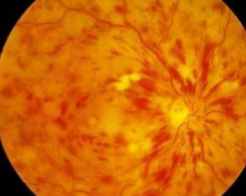
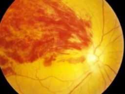
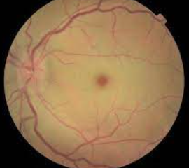
Over 28,000 cases of retinal detachment occur every year.
Retinal detachment is sight-threatening retinal disease and occurs when the retina separates from the back of your eye, causing loss of vision that can be partial or total, depending on how much of the retina is detached.
The annual incidence of retinal detachments is approximately 12.5 cases per 100,000 per year, or about 28,000 cases per year in the US.
If you suffer any sudden vision changes, immediately contact an eye doctor near you.
What causes retinal detachment?
The round shape of the eye is maintained by a gel, called the vitreous. The vitreous is attached to the back of the eye, the retina, and provides a soft cushion for the retina.
However, the vitreous gel can sometimes become detached from the retina and this serious retinal disease happens when the vitreous gel pulls away sufficiently to tear off a piece of the retina. An influx of fluid through the tear may further lift and detach the retina.
Signs and symptoms of retinal detachment
While there is no pain associated with retinal detachment, it’s important to be aware of signs that the retina has become detached.
Symptoms include:
- Sudden blurred vision
- Flashes of light that appear in the side of your vision
- Suddenly seeing many floaters that look like lines, specks or cobwebs in your field of vision
- Partial vision loss, such as a shadow in your peripheral vision or a gray curtain covering part of your vision
If you’ve experienced any of the above symptoms, immediately contact an eye doctor near you.
SEE RELATED: Retinal Holes and Tears
Who is at risk for retinal detachment?
Risk factors for retinal detachment include:
- A family history of retinal detachment
- Over 50 years old
- Diabetes mellitus
- Having a serious eye injury in the past
- Previous cataract, glaucoma, or other eye surgery
- High level of myopia (near-sighted)
- Prior history of retinal detachment
- Taking glaucoma medications
- Trauma to your eye
How to treat retinal detachment
In most cases, surgery is necessary to repair all retinal disease and especially a detached retina.
Some types of retinal surgery procedures include:
1.Vitrectomy
A vitrectomy is used for larger tears. Small tools are used to remove abnormal vascular or scar tissue and vitreous, a gel-like fluid from your retina. The vitreous will be replaced with an air, gas, or oil bubble. The bubble pushes the retina back into place so it can heal properly.
2. Scleral buckle
A more severe detachment requires eye surgery in a hospital. Scleral buckling involves placing a band of rubber or soft plastic onto the outside of your eyeball. It gently presses the eye inward, helping the detached retina heal against the eyewall. The scleral buckle is not seen on the eye and is usually left on the eye permanently.
3. Photocoagulation
This procedure is used if there is a hole or tear in your retina but your retina is still attached. The eye doctor uses a laser that burns around the tear site. The resulting scarring binds your retina to the back of your eye.
4. Cryopexy
Cryopexy is freezing with intense cold. For this treatment, your eye doctor will apply a freezing probe outside your eye in the area over the retinal tear. The resulting scarring helps hold the retina in place.
5. Retinopexy
A pneumatic retinopexy is used to repair minor detachments. For this procedure, a gas bubble will be placed in your eye to help your retina move back into place up against the wall of your eye. Once your retina is back in place, your eye doctor will use a laser or freezing probe to seal the holes.
While there’s no way to prevent retinal detachment, you can take steps to reduce your risks of suffering from retinal detachment.
Seeing your eye doctor frequently, such as yearly eye exams, especially if you have risks for retinal diseases, is important to avoid vision loss.
LEARN MORE: Guide to Retinal Disease
Schedule an emergency eye exam with an eye doctor near you so you can avoid any retinal problems and maintain a life of good eye health and clear vision.


